Table Of Contents
1. Introduction
2. What Is Image Analysis?
3. What Are The Important Steps Of Image Analysis?
4. What Are The Applications Of Image Analysis?
5. What Are The Challenges That Might Occur While Performing Image Analysis?
6. What Are The Image Analysis Software Tools Used For Microscopy?
7. Conclusion
8. Key Takeaways
Introduction
Image analysis is a field of study and technology that is especially dedicated to the evaluation and interpretation of visual information. It includes the usage of computational techniques in order to extract meaningful data from images, whether they are satellite pictures, photographs, medical scans or any other visual data.
Image analysis plays an important part in various applications that we are going to discuss in this article today. We are also going to learn about the steps involved in the image analysis process and much more. Let's begin with understanding the meaning of image analysis!
What Is Image Analysis?
Image analysis, also known as image processing, is a field of computer science and engineering that specifically focuses on the extraction of valuable data from digital images. It typically encompasses the utilization of numerous methods and algorithms in order to analyze, manipulate and interpret images in a digital format. Image analysis plays a vital role in different fields, which includes computer vision, remote sensing, medical imaging, quality control and much more.
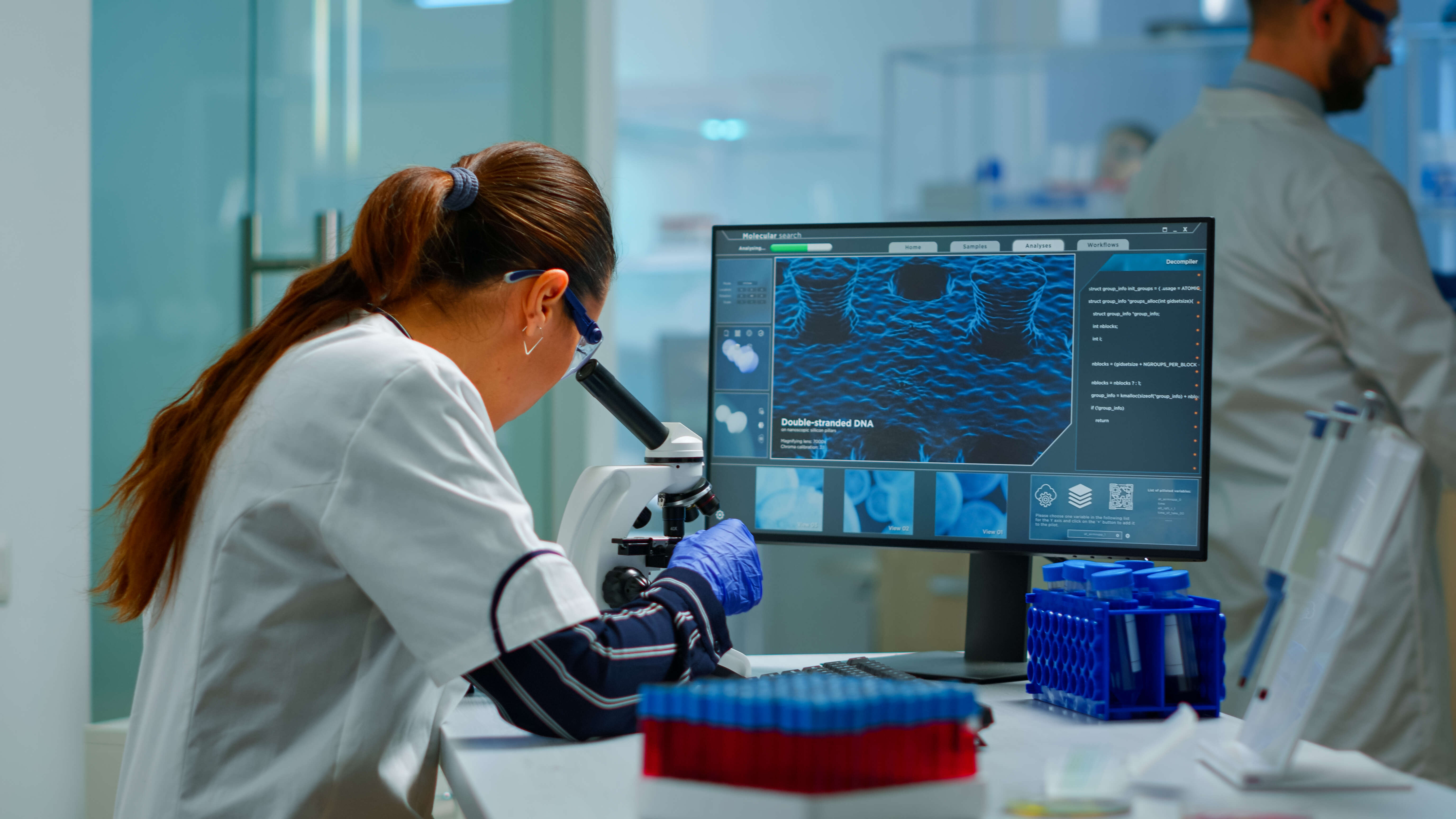
Types Of Image Processing:
a) Visualization: Detecting specific objects that are not clearly visible in the picture.
b) Recognition: This means distinguishing or identifying the components in the image.
c) Sharpening & Restoration: Developing an improved picture from the original picture.
d) Pattern Recognition: Estimating the numerous patterns of the objects present in the image.
e) Retrieval: Browse as well as look for images from a huge database that contains a high number of images and is identical to the original image.
What Are The Important Steps Of Image Analysis?
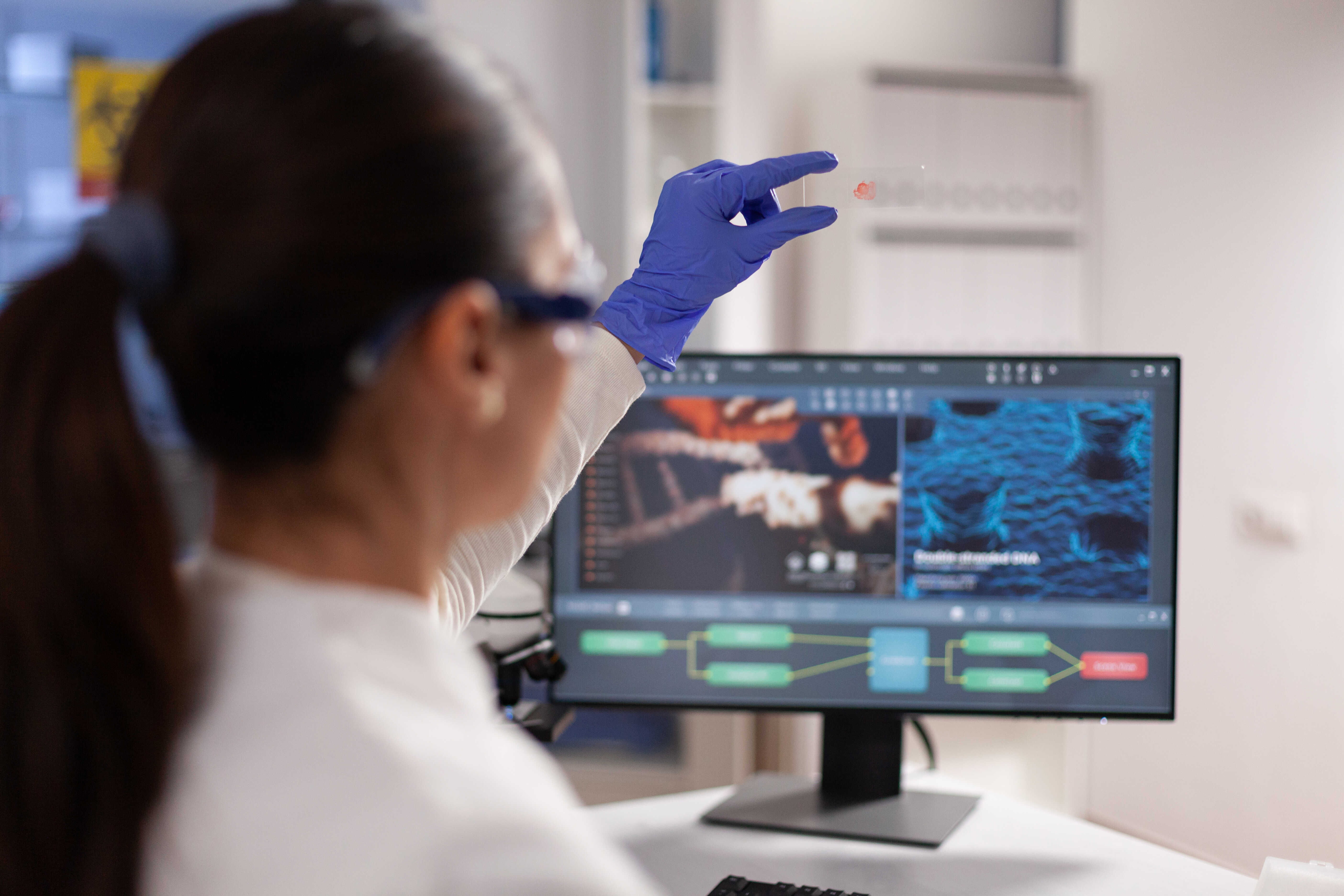
a) Image Acquisition — The process of image analysis starts with the acquisition of digital pictures. All these images may come from different sources, such as cameras, medical devices, scanners or satellites. The quality of such images can have a significant impact on the subsequent analysis.
b) Image Pre-Processing — Before the analysis of the image takes place, it becomes essential to pre-process all the images. This might encompass work such as enhancement of the image, reduction of noise and correction of certain distortions or artifacts. Pre-processing of images intends to improve the quality of the data for further examination.
c) Image Segmentation — Image segmentation includes tasks such as partitioning an image into meaningful areas or entities. It is considered to be an essential step in image analysis because it tends to isolate the areas of interest from the environment. Image segmentation is repeatedly utilized for the detection and tracking of images.
d) Feature Extraction — After the segmentation of the images is done, some of the relevant characteristics or features are extracted from these areas. Features include colour, shape, size, texture or any other measurable attribute that helps in distinguishing one element from the other.
e) Pattern Recognition — The process of image analysis generally involves pattern recognition methods. All these techniques are utilized to extract features in order to identify and classify objects, find anomalies or make informed decisions on the basis of the image contents.
f) Data Analysis And Interpretation — Now the extracted data is utilized to draw conclusions, make important predictions or obtain insights that are associated with the original image. All these steps might involve machine learning, statistical analysis, or any other analytical techniques.
What Are The Applications Of Image Analysis?
Image analysis has a wide range of applications, some of which are mentioned below:
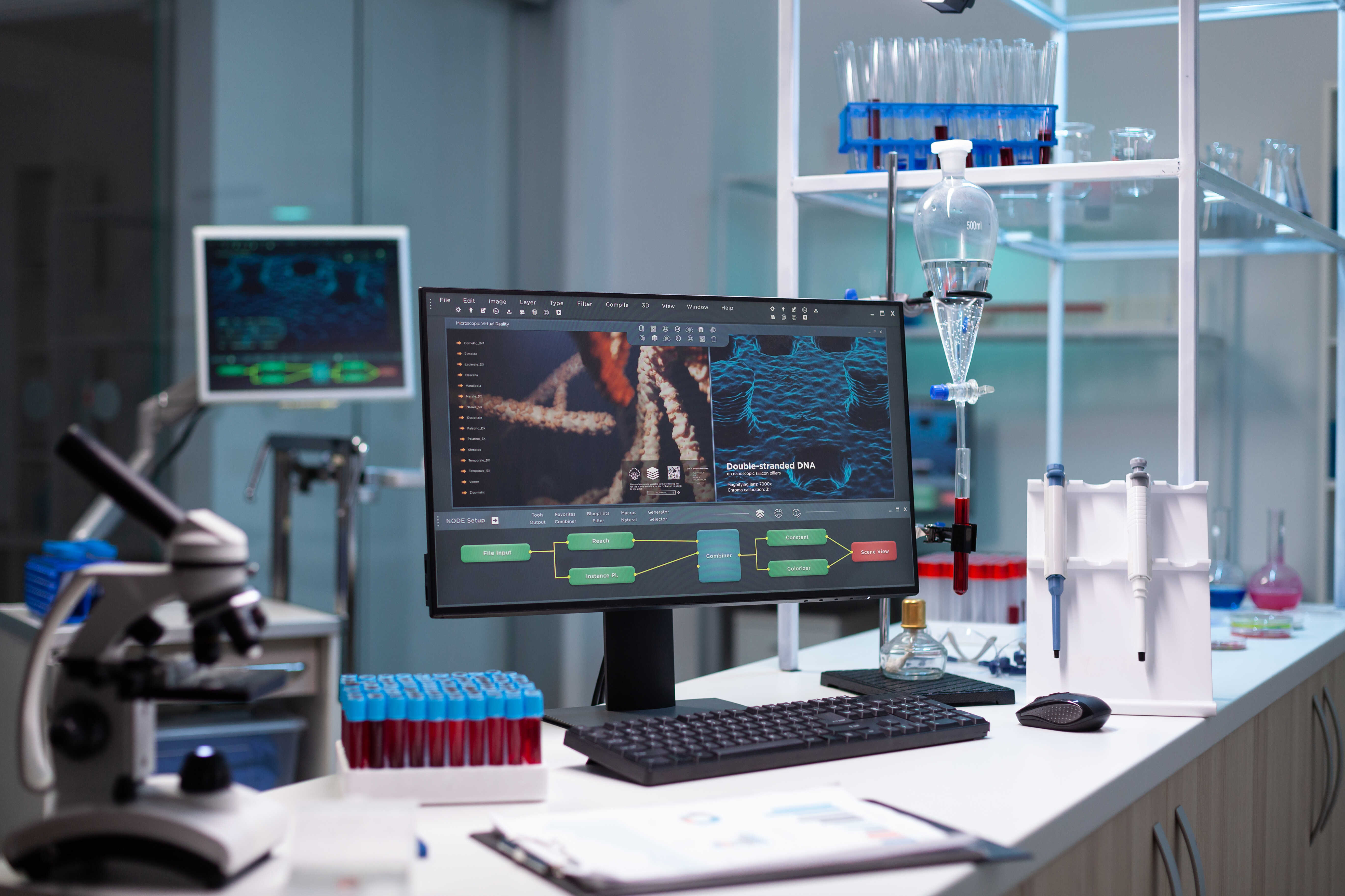
1) Medical Imaging:
Image analysis is utilized in medical imaging to help in the diagnosis of numerous diseases and conditions. It assists in detecting fractures, tumours, and certain abnormalities in MRIs, X-rays and CT scans. Additionally, it is also utilized for analyzing tissue samples in order to detect ailments or examine their progression.
2) Object Recognition And Tracking:
In the field of robotics and autonomous vehicles, image analysis is utilized to acknowledge, track various objects and navigate the environment.
3) Astronomy:
Image analysis or processing is even utilized to process astronomical images and determine celestial objects like stars, galaxies, and planets.
4) Quality Control And Manufacturing:
This specific method is utilized to examine products on manufacturing lines so that defects or irregularities can be detected and ensure quality control. In the semiconductor industry, image analysis is utilized to check for deficiencies in microchips.
5) Security And Surveillance:
Image analysis is employed in surveillance systems to detect and keep track of intruders, monitor traffic, and identify faces for security objectives.
What Are The Challenges That Might Occur While Performing Image Analysis?
The image analysis process can be a challenging task because of the variations in lighting, complex backgrounds, image noise and the requirement to develop robust algorithms that really work in real-world scenarios. In addition, huge datasets and computational strengths are generally needed for deep learning-based image analysis.
Image analysis, the process of extracting meaningful information or features from digital images, presents several challenges, including:
1) Variability In The Quality Of The Image — The images might vary in terms of lighting, focus, noise and resolution. Evaluating the images that are of poor quality can result in incorrect outcomes.
2) Lighting And Shadows — When it comes to the variations in lighting and shadows, they can have an effect on the appearance of certain objects and might even complicate their identification and analysis.
3) Color Variability — Images might also contain specific objects that have different colours and colour patterns, and this can bring in a lot of difficulty in segmentation and classification.
4) Complexity Of Images — Natural scenes and elements can be highly complex and identifying as well as interpreting all these elements in occluded or cluttered environments is quite challenging.
5) Scale And Perspective — There may be some images that may also contain objects or regions at different scales and viewpoints, making it tricky enough in order to extract features unfailingly.
6) Data Annotation — The process of image analysis usually needs labelled data for training machine learning models. Annotating images with ground reality data can be time-consuming and pricey.
7) Computational Resources — Processing and analyzing huge numbers of high-resolution images might come across as computationally intensive, mandating substantial computing strength as well as memory.
What Are The Image Analysis Software Tools Used For Microscopy?
Here are some of the best image analysis software tools utilized for microscopy:
1) ipvPClass — ipvPClass stands as a sophisticated evolution of the Particle Size Analyzer, delivering a remarkable combination of speed, convenience, and accuracy in particle analysis. This advanced system, known as ipvPClass, is a comprehensive Microscopic Image Analysis platform. It seamlessly integrates a cutting-edge high-resolution camera with dedicated software, making it adaptable to a wide range of microscopes. Its primary purpose is to facilitate the identification of particulate solids within the microstructure of various samples.
2. ImageJ/FIJI — ImageJ is a highly popular, open-source software which is specifically designed for image processing as well as analysis. It is essential to be proficient at utilizing ImageJ for image analysis. Using this, you can do a few simple things such as crop, label or modify the temperature of the images. It also has the capability to manage 3D stacks of confocal microscopy images and conduct complicated quantitative analysis.
Similarly, FIJI is a distribution of ImageJ that comes with multiple pre-installed plugins and a user-friendly interface. It provides a wide range of image analysis tools, is highly extensible with plugins, and can manage different image formats. It is incredibly fitted for biological and biomedical microscopy.
3. CellProfiler — CellProfiler is an open-source software that is created for high-throughput image analysis. It is well-suited for cellular as well as subcellular analysis and is typically utilized in cell biology and histopathology. CeProfiler comes with a user-friendly interface for building analysis pipelines, and it provides a comprehensive range of image analysis modules. This particular software is greatly useful for analyzing cell populations.
4. L-Measure — This one is another software tool designed for the analysis of neural structures in two-dimensional (2D) as well as three-dimensional (3D) images. It is generally utilized in the field of neuroanatomy and neuroscience in order to quantify and measure different morphological and architectural features of neurons, such as axons and dendritic trees. L-Measure delivers a systematic way to extract and examine varied quantitative parameters from digital reconstructions of neuronal structures.
5. Volume Integration and Alignment System (VIAS) — VIAS facilitates the consolidation of multiple confocal microscopy image stacks into a unified 3D image dataset. Developed by the same team responsible for creating Neuronstudio, this software boasts a comparable user-friendly interface and provides a comprehensive step-by-step online manual.
This tool offers the convenience of real-time interaction with the image stacks, permitting users to effortlessly reposition them until they achieve perfect alignment. Additionally, an automated alignment feature is available to fine-tune the alignment for a seamless end result.
In cases where image stacks have varying depths, VIAS allows for straightforward alignment in the z-dimension. Upon completion, the resulting dataset can be exported as a .tif file for utilization in ImageJ.
6. QuPath — QuPath is also an open-source software developed particularly for the analysis of digital pathology images. It is favourable for performing tasks such as tissue segmentation and whole-slide image analysis. It provides tools for tissue detection, cell segmentation, and beyond. It is widely utilized in pathology research and diagnostics.
7. Icy — Icy, is an open-source bioimaging software that offers tools for processing and analyzing a vast range of microscopy information, which includes time-lapse and fluorescence and imaging. It sustains different types of image formats and provides a plugin system for expanding its capabilities. It is commonly utilized in life sciences.
8. ImagePy — ImagePy is a widely known open-source, cross-platform image processing framework that provides a number of tools for image analysis as well as visualization. The best part about this particular tool is that it is extensible and can be customized with various plugins. It is not just restricted to microscopy and can be utilized for general image processing.
Conclusion
Image analysis and its associated software tools have been known to revolutionize different fields by enabling the extraction of meaningful insights from visual data. This robust technology has a wide range of applications, varying from healthcare to agriculture, from surveillance to art restoration.
With the advancement of machine learning and deep learning, image analysis software has become increasingly sophisticated, permitting more accurate and versatile examinations. It has not only enhanced the velocity and efficiency of decision-making procedures but also unlocked the door to automation and data-driven discoveries.
However, if you wish to learn more about this, then you must visit our website ImageProvision and in case of any queries, contact us anytime!
Key Takeaways
- Image analysis pertains to extracting valuable data or input from digital images.
- Image analysis software makes use of algorithms to process and interpret images.
- Key applications of image processing techniques include medical imaging, remote sensing, and quality control.
- Machine learning and deep learning enhance the capabilities of image analysis.
- Software tools such as these are popular for image analysis techniques.
- Image segmentation, feature extraction, and object recognition are common steps in the process of image analysis.
- Image analysis helps in decision-making, automation, and data-driven insights.


